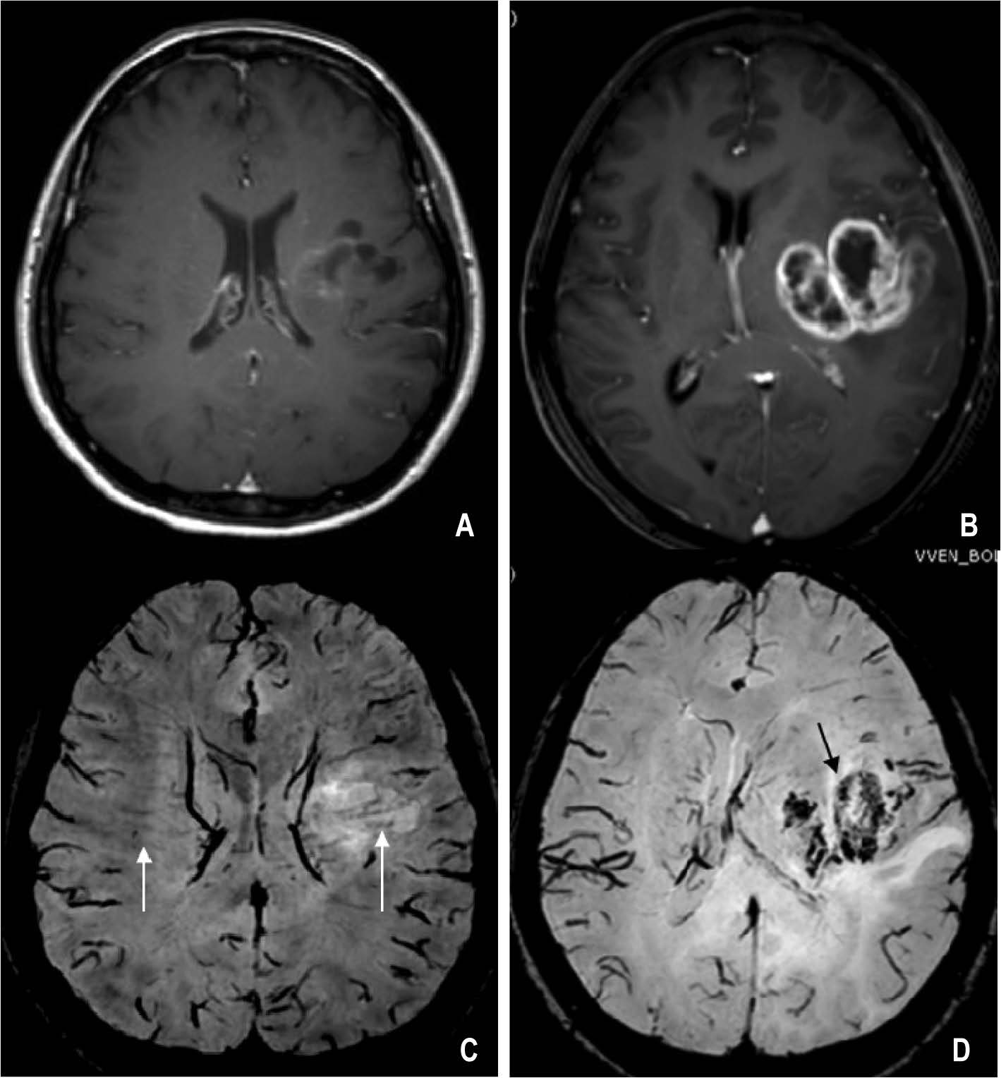
(A, B) Gd-T1 W, (C, D) SWI: comparing between multiple sclerosis (A, C) and glioblastoma multiforme (B, D). There is enhancement at the periphery of both lesions. On SWI, the transmedullary vein is seen passing the MS lesion (arrows in C). In glioblastoma multiforme the normal vein was destroyed and replaced by tumor microvasculature (arrow in D).
Figure 2:
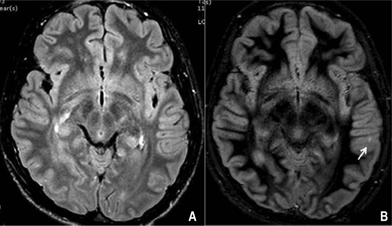
Double inversion recovery (DIR) in multiple sclerosis: (A) FLAIR, (B) DIR show lesion in the gray matter of cerebral cortex on DIR (arrow in B) which is not seen on FLAIR.
Figure 3:
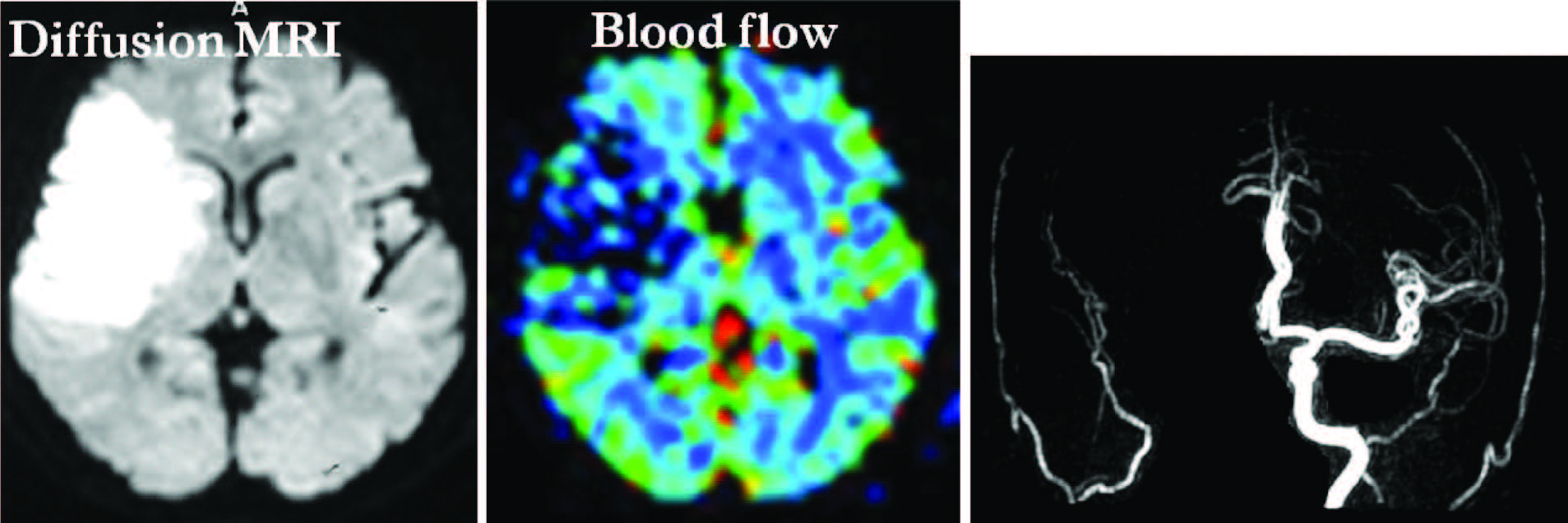
Acute infarction due to occlusion of right internal carotid artery showing lesion on DWI corresponding to perfusion MRI (blood flow) or “diffusion/perfusion match”. It means totally infarct without penumbra of the ischemic area and no advantage to apply thrombolytic agent.
Figure 4:
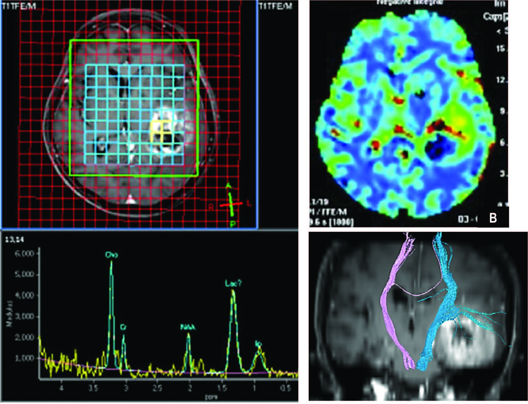
Hight gradeglioma (A) MRS, (B) p MRI (CBV), (C) DTI. MRS shows definitely increased Cho, Lac and Lip peaks in brain tumor. The NAA is markedly decreased. pMRI demonstrates increased blood volume at the periphery (arrow in B) and no blood supply of the central necrosis. DTI shows the relationship between the tumor and the ipsilateral corticospinal tract.
Figure 5:
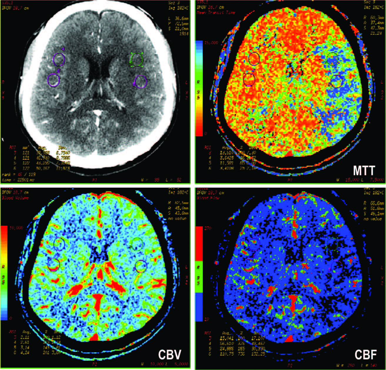
CT perfusion of a patient presented with sudden right sided weakness. There is no detectable abnormality on CECT. The color map of MTT and CBF show the ischemia in left brain (left MCA territory). The CBV does not show definite abnormality.
Figure 6:
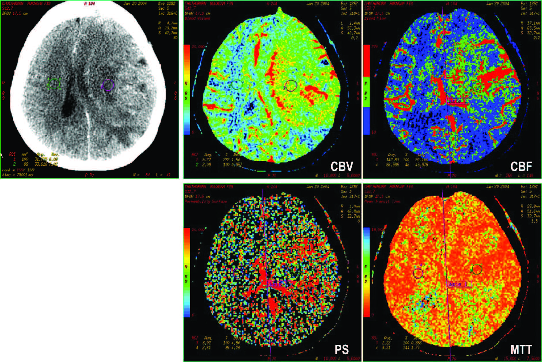
CT perfusion of a patient diagnosed glioblastoma multiforme, status post craniotomy. The surgical bed of left cerebrum shows definitely increased CBV, CBF. The icreased perme- ability surface index (PS) indicates recurrent brain tumor.
Figure 7:
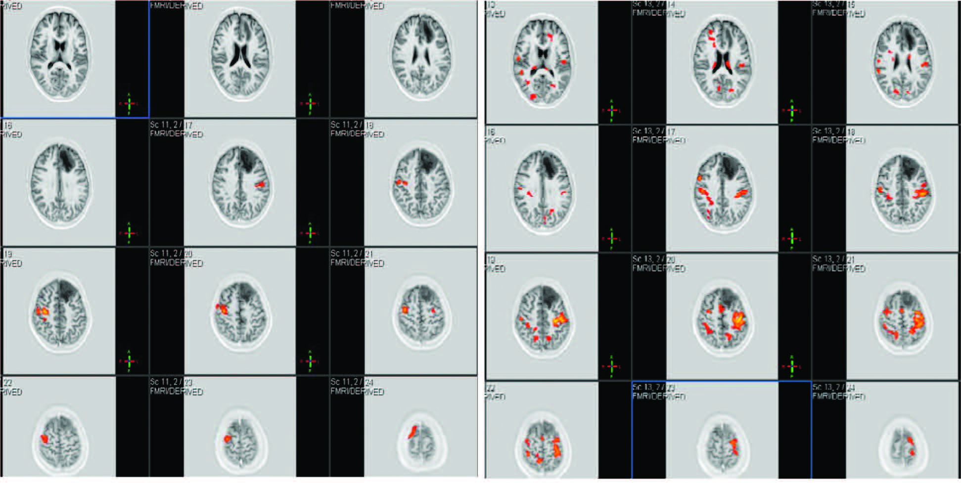
Functional MRI shows areas of stimulated brain during left hand movement (A), and right hand movement (B). There is a brain tumor in the left frontal lobe.