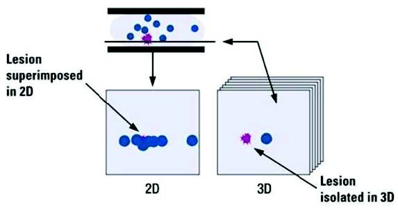
Overlapping structures hides lesions in two dimensional (2D) that can be clearly seen in a three dimensional (3D) cross sectional slice.
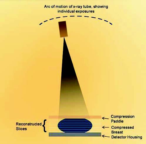
Figure 2:
Schematic shows principle of operation of tomosynthesis system.
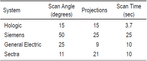
Table 1:
Scan parameters used in some tomosynthesis systems.
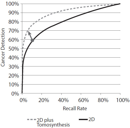
Figure 3:
The diagonal arrow shows how an individual’s cancer detection rate can be improved and their recall rate reduced using a technology that has a higher ROC curve.
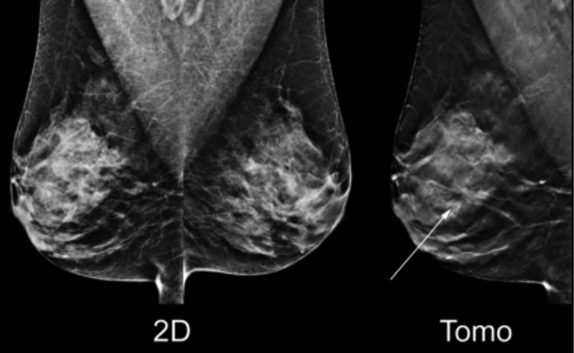
Figure 4:
Increased cancer detection: the tomosynthesis reconstructed slice shown on the right reveals a definitive spiculated mass that is only faintly revealed in the 2D image shown on the left. (Diagnosis: Invasive Ductal Carcinoma)
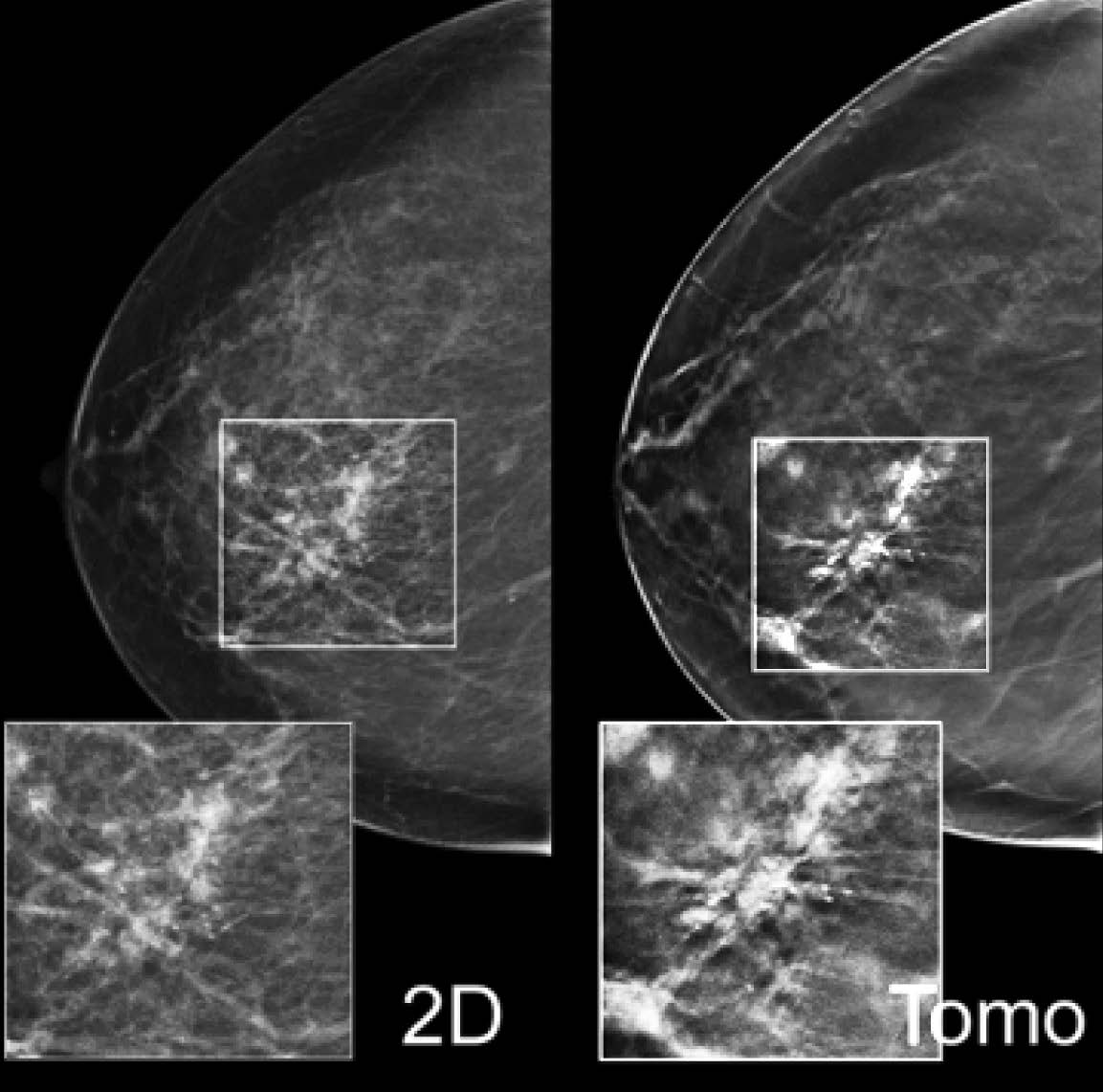
Figure 5:
Added value for calcifications: the 2D mammogram on the left shows right medial microcalcifications. The tomosynthesis reconstructed slice on the right illustrates the associated architectural distortion only revealed on the CC tomosynthesis image and not shown on the mammogram. (Diagnosis: Ductal Carcinoma In-situ/High Grade)
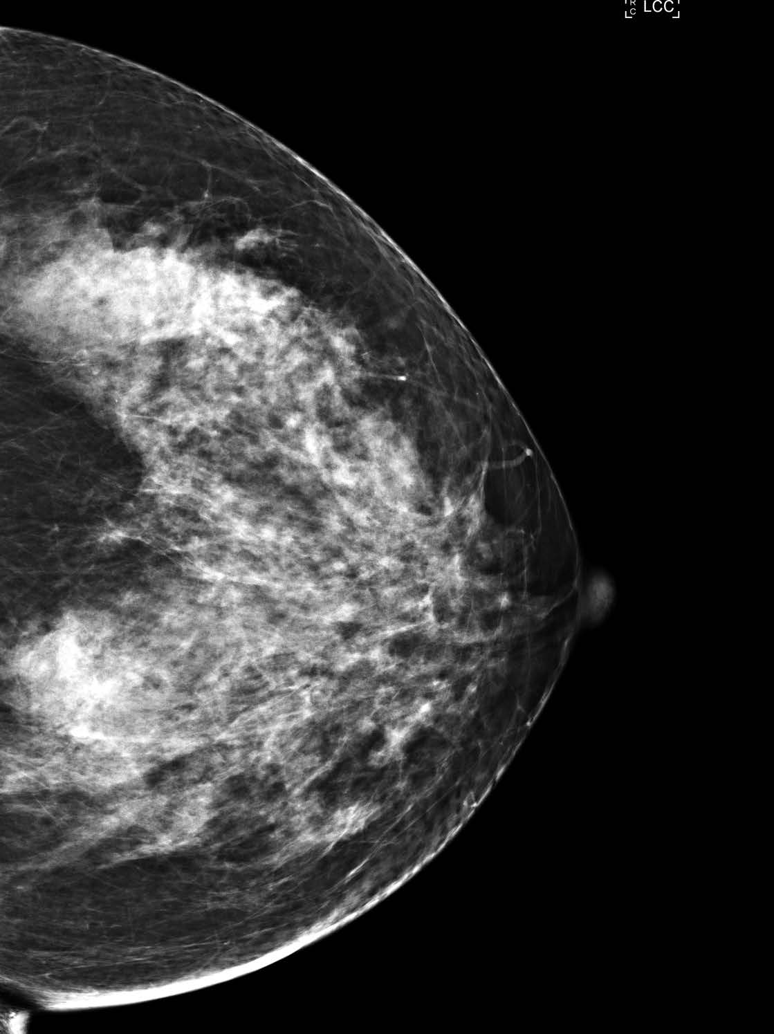
Figure 6:
The 2D mammogram reveals a heterogeneous dense breast with a craniocaudal (CC) view.
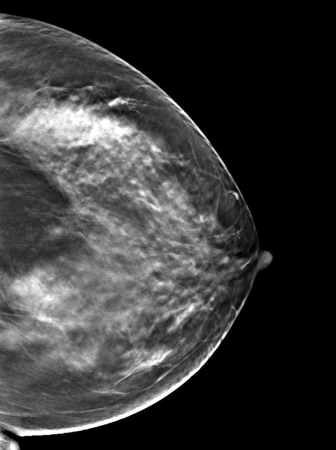
Figure 7:
The 3D breast tomosynthesis shows a well defined mass on the upper inner quadrant; the size is 16x18mm (arrow).
Figure 8:
The ultrasound is inconclusive in identifying the mass