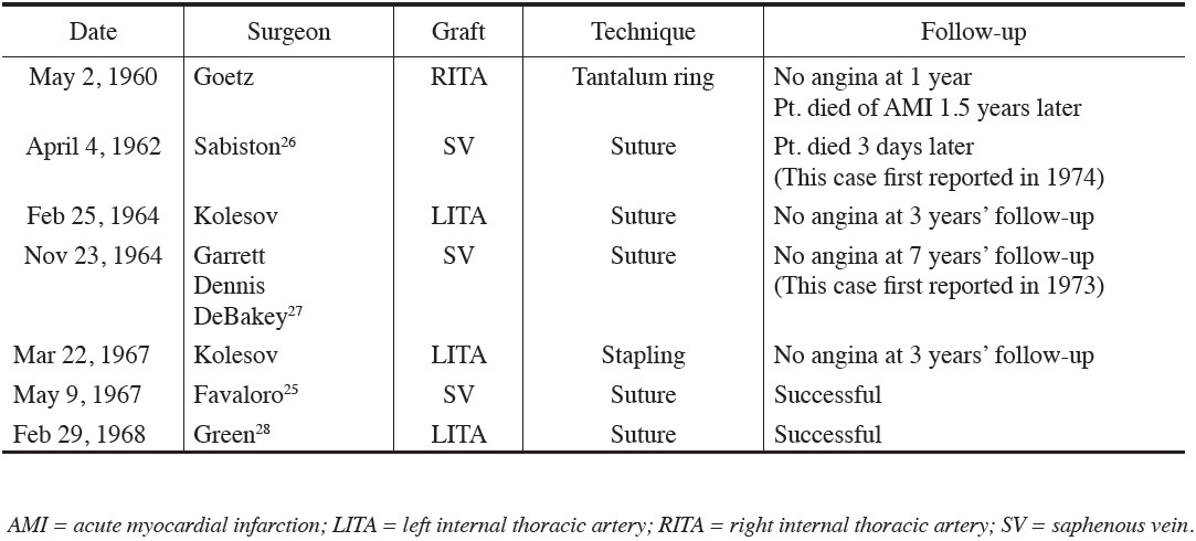
The First Clinical Coronary Artery Bypass Operations
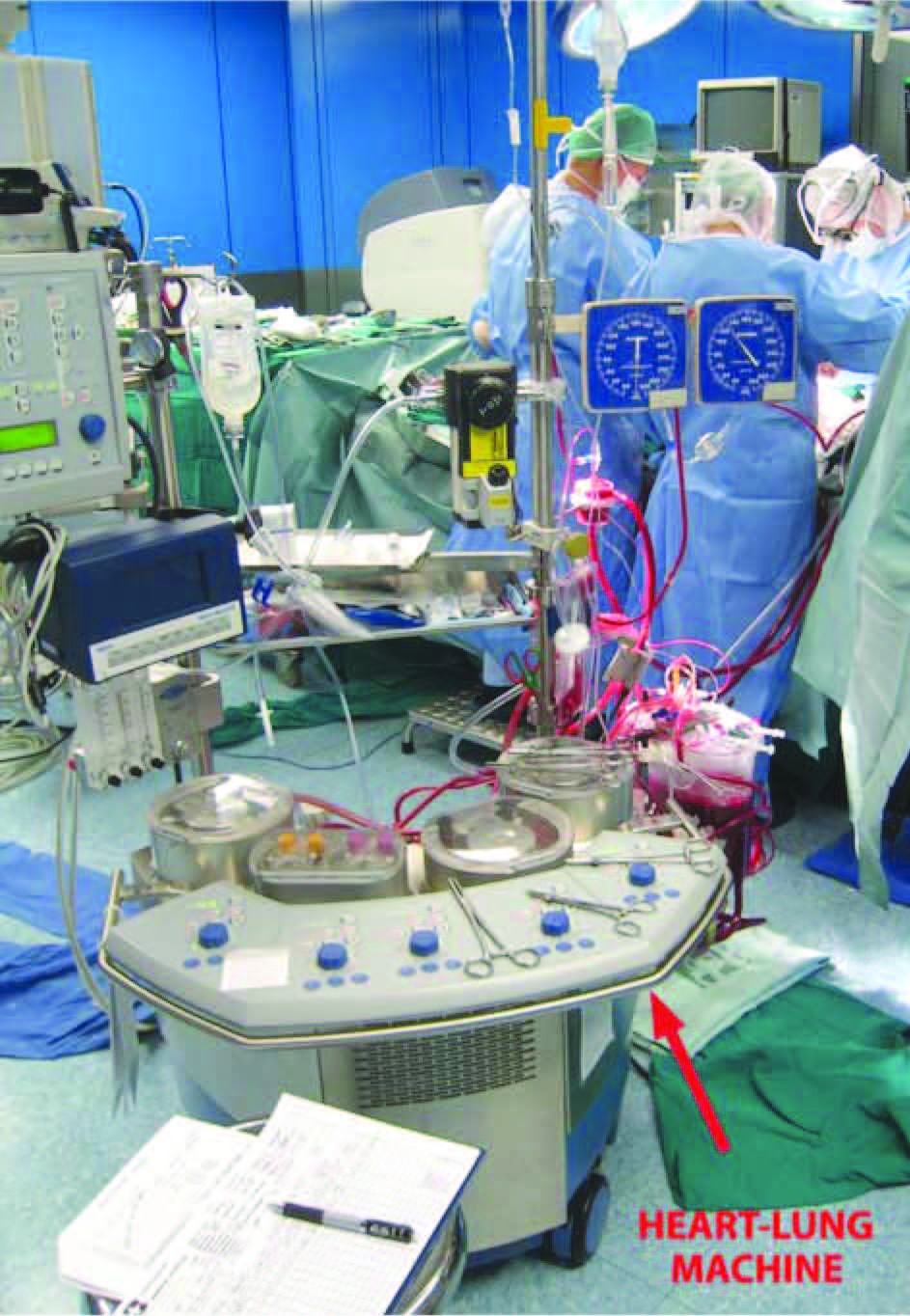
Figure 1:
Heart-Lung Machine at the Bangkok Heart Hospital
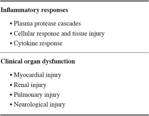
Table 2:
The side effects of cardiopulmonary bypass
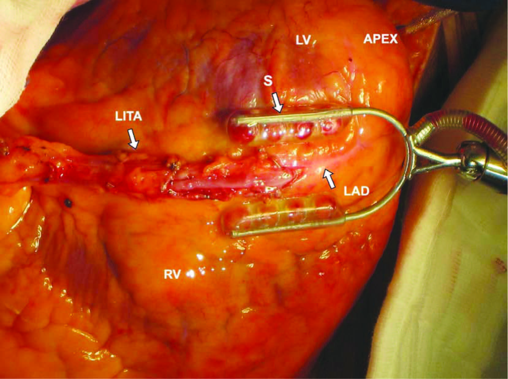
Figure 2:
The left internal thoracic artery was anastomosed to the left anterior descending artery with off-pump technique. Stabilizing arm with suction foot is on the area of anastomosis of the left anterior descending artery.
LV = Left ventricle, S = Stabilizer, LITA =Left internal thoracic artery,
LAD = Left anterior descending artery, RV = Right ventricle
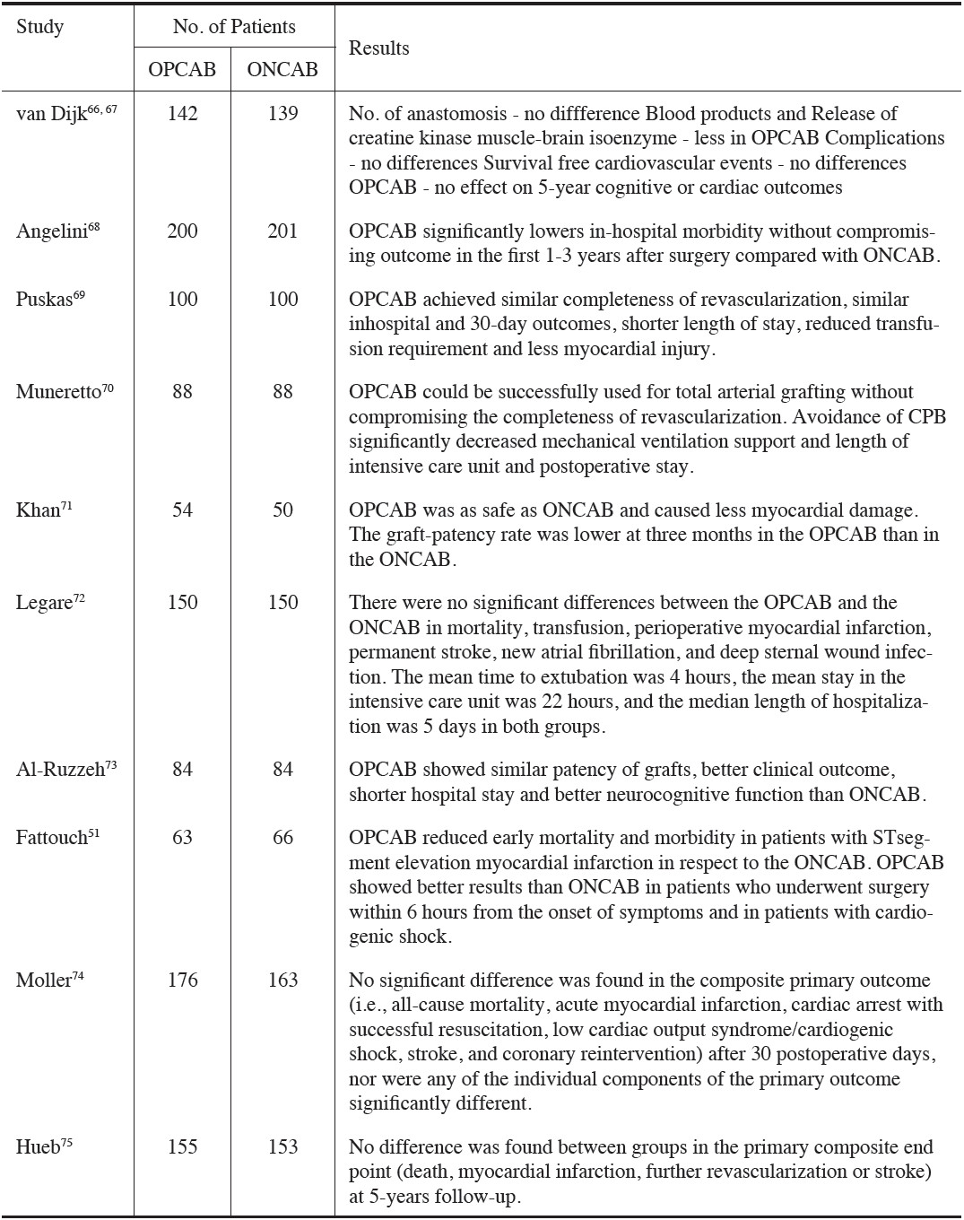
Table 3:
Summary of the randomized controlled trials comparing off-pump (OPCAB) and on-pump (ONCAB) surgery
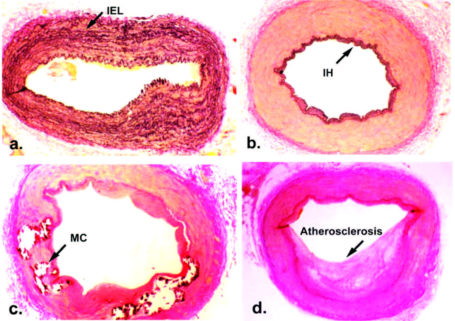
Figure 3:
Histopathology of the internal thoracic artery (ITA) and radial artery (RA), (a.) The ITA demonstrates internal elastic lamina (IEL). [Verhoeff Van Gieson’s elastin ´ 20 (original magnification) ] (b.) The RA demonstrates intimal hyperplasia (IH). [Verhoeff Van Gieson’s elastin ´ 20 (original magnification) ] (c.) The RA demon- strates medial calcification (MC) in the media of the arterial wall. [Haematoxylin-eosin´25 (original magnifica- tion) ] (d) The RA demonstrates atherosclesosis. [Haematoxylin-eosin´25 (original magnification) ]
ITA = internal thoracic artery; RA = radial artery; IEL = internal elastic lamina; IH = intimal hyperplasia; MC = medial calcification
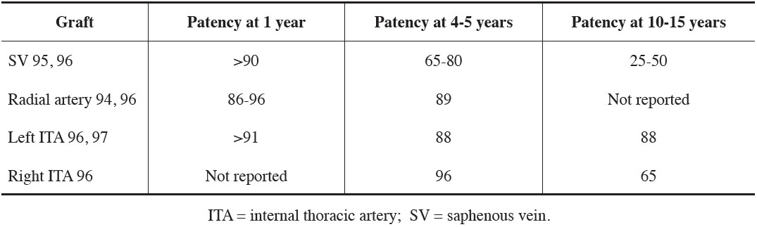
Table 4:
Graft patency after coronary artery bypass grafting (%)
Figure 4:
256 slice computed tomography volume-rendering image in the left anterior oblique projection. (a.) the radial artery (aortocoronary graft) to the obtused marginal branch of the left circumflex artery (b.) the radial artery (T graft from the left internal thoracic artery) to the obtused marginal branch of the left circumflex artery.
T = T graft LV = Left ventricle
AO = Aorta LITA = Left internal thoracic artery
AC = Aortocoronary graft LAD = Left anterior descending artery
RV = Right ventricle
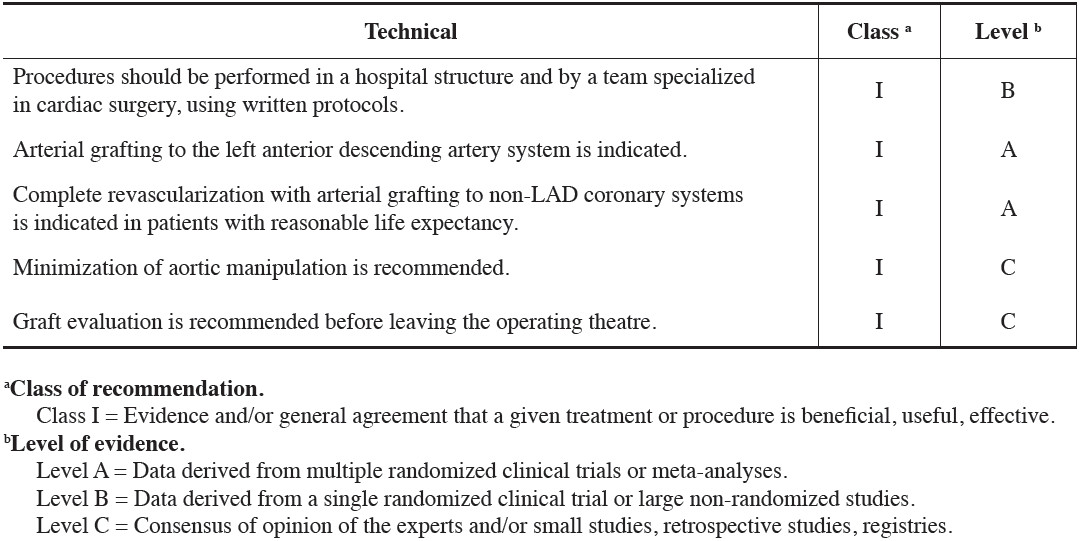
Table 5:
Technical recommendations for coronary artery bypass grafting