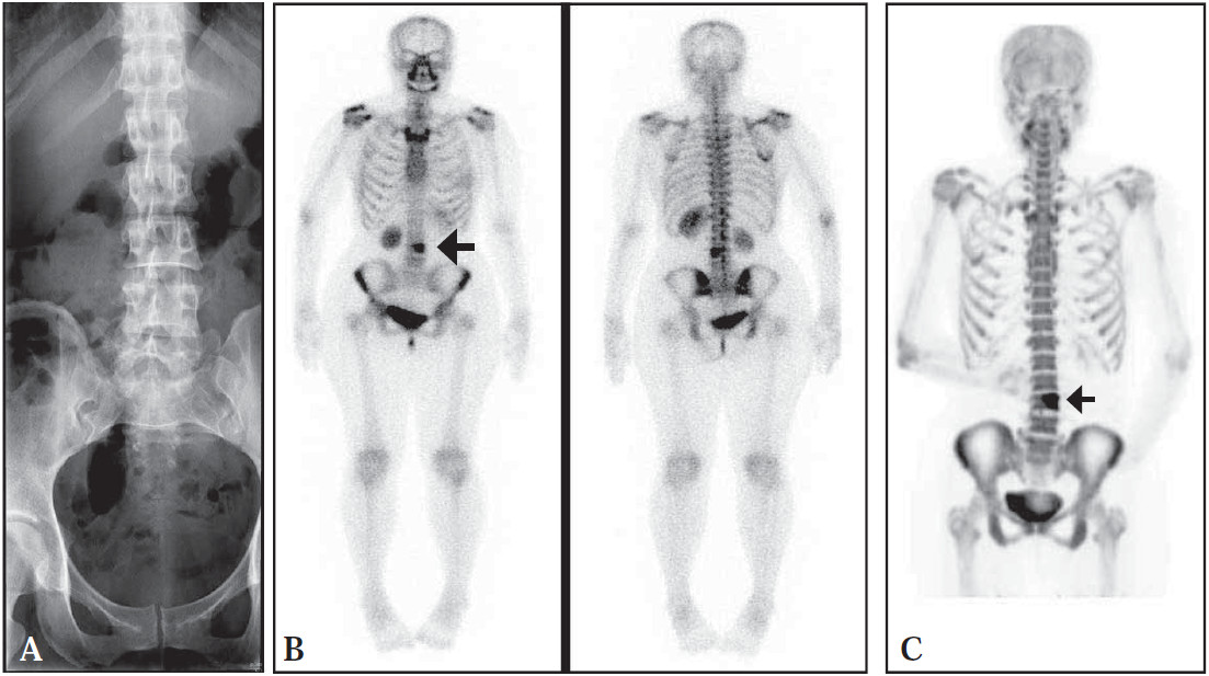
Figure 1: A. Radiograph of lumbar spine shows unremarkable findings. B. Anterior and posterior whole body bone scan (Tc-99m MDP) show increased radioactivity uptake at left aspect of L4 (arrow).
C. Anterior whole body PET/CT (F-18) shows increased radioactivity uptake at left aspect of L4 (arrow).
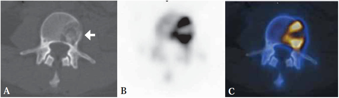
Figure 2: A. CT part of PET/CT image shows osseous destruction at vertebral body of L4 (arrow) (Same patient as fig. 1C) B. PET scan shows abnormal activity at the corresponding osseous destruction and C. Fusion PET/CT of the osseous metastasis.