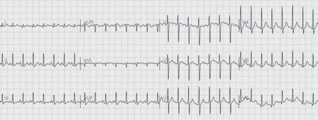
Figure 1: Electrocardiogram on admission shows sinus tachycardia.
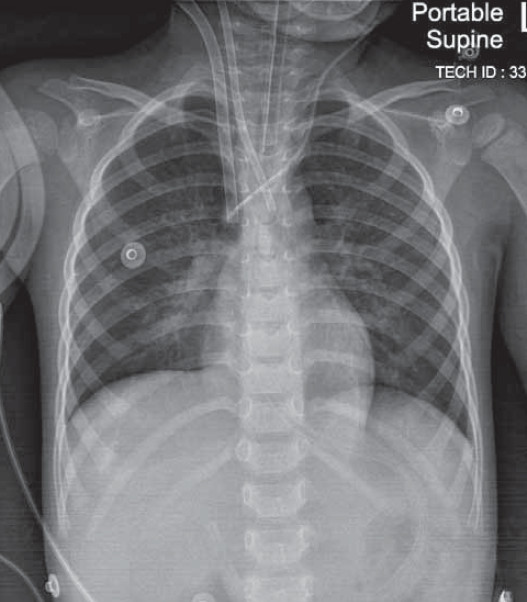
Figure 2A: On January 20, 2013 at 15:21, a chest x-ray AP supine position reveals early pumonary edema. Heart appeared normal. The tip of catheter was in SVC, endobroncheal tube is in place.
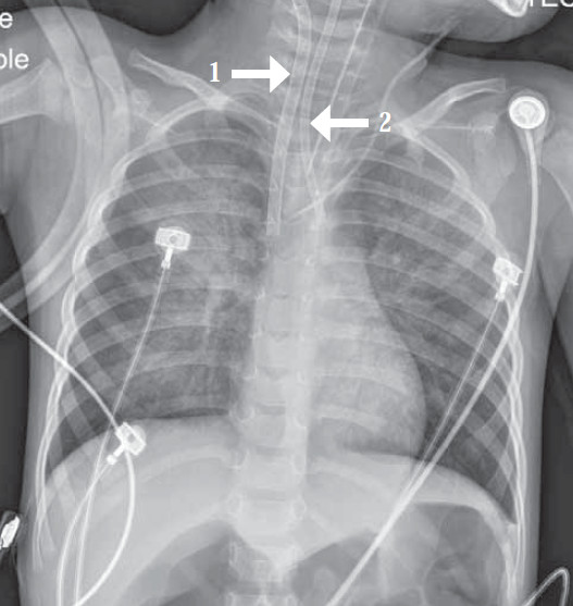
Figure 2B: On January 23, 2013 at 21:41, a chest x-ray AP supine position and ECMO venous to arterial connection. The tip of catheter 1 is in SVC. The tip of catheter 2 is in aortic arch. Progressive pulmonary edema developed.
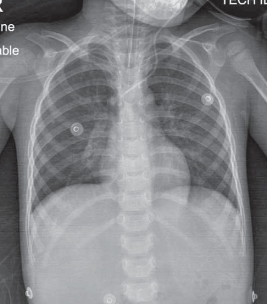
Figure 2C: On January 24, 2013 at 15:38, a chest x-ray AP supine position reveals improvement of pulmonary edema.
Figure 2D: On January 27, 2013 at 5:30, a chest x-ray AP supine position and post removal ECMO reveals heart and lung appear normal.
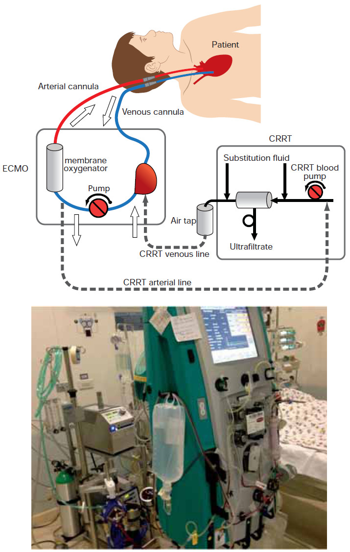
Figure 3: Diagram of venous-arterial connection with continuous renal replacement therapy (CRRT) incorporated into the circuit.