Electronic ISSN 2287-0237
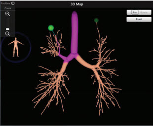
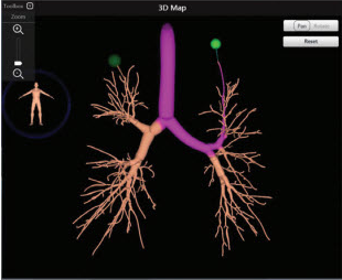
Suspicious pulmonary nodules have become a more common finding since the advent of CT scans as a screening method for lung cancer. The challenge is to find a safer, practical and more accurate way to diagnose peripheral nodules and lesions while minimizing harm to the patient. Electromagnetic Navigation Bronchoscopy (ENB) is a cutting edge technology that enables pulmonologists to precisely target small and difficult to reach peripheral pulmonary nodules by using technology similar to a global positioning system (GPS).This study describes our experience in using ENB with Endobronchial ultrasound (EBUS) as a diagnostic tool on patients suspected of lung malignancy.
A retrospective analysis was conducted for 33 patients between November 2014 and November 2015 at our institution. Yield was determined by attaining final diagnosis from tissue analysis of every patient who underwent the ENB and EBUS procedure for suspected lung malignancy. Patients with no definite diagnosis were referred to surgery for further examination. Intraoperative and postoperative complications were recorded.
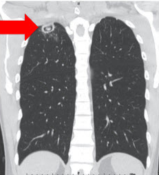
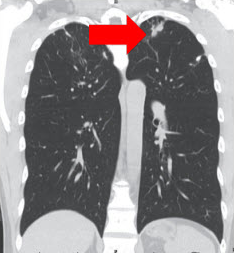
The ENB and EBUS procedure was completed in all 33 subjects on lung nodules measuring from 2 to 108mm. The patients were 67% (22) male, 33% (11) female, 88% (29) Asian, with a mean age of 61 years old. Lung nodules were located in the upper lobe (52%), lower lobe (43%) and middle lobe (5%). Lymph node involvement was present in 33% of cases. Of the patients in the study, 26 cases (79%) were accurately diagnosed using ENB and EBUS, 6 cases (18%) were referred to surgery and 1 patient (3%) refused further investigation. No bleeding or pneumothorax complications were reported.
ENB is an effective, accurate and a novel method that provides real time directions toward lung lesions and nodules. This tool allows flexibility that aids biopsy of hard to reach areas, and attains adequate tissue samples. This instrument compliments other bronchoscopic methods such as EBUS.
10.31524/bkkmedj.2017.02.005