Electronic ISSN 2287-0237
A 51-year-old man presented with chronic diarrhea since 7-8 years. The physical examination was within normal limits. The laboratory tests were unremarkable.
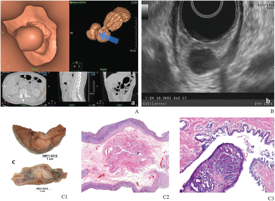
Virtual gastroscopy (Figure A) revealed a submucosal mass at the antrum on greater curvature of stomach 1.4 x 8 cms. EUS (Figure B) showed heterogenic hypoechogenic lesion at 3rd and 4th layers of gastric wall. Removal of gastric mass through gastroscopy was performed. The gross examination demonstrated a small submucosal protruding nodule 1.0 cm. in diameter but, no evidence of mucosal ulceration. Serial sections showed no definite connection between the nodule and gastric mucosa, nor evidence of ulceration at the mucosa. The microscopic section (Figure C1-3) showed pancreatic tissue composed of a mixture of acinar tissue and ductal epithelial element, the endocrine element was not clearly seen.
Gastric aberrant pancreas is an unusual condition, it is only reported sporadically. The findings revealed submucosal mass without ulceration. Upper GI study and gastroscopy are well established methods, however virtual gastroscopy may well be as demonstrative as other established modalities. EUS showed heterogenic hypoechogenic mass in 3rd and 4th layers of stomach as shown on the previous literature.1


