3D reconstruction, Liver mass, hepatoma, cholangiocarcinoma
DOI
10.31524/bkkmedj.2012.02.014
MEDIA
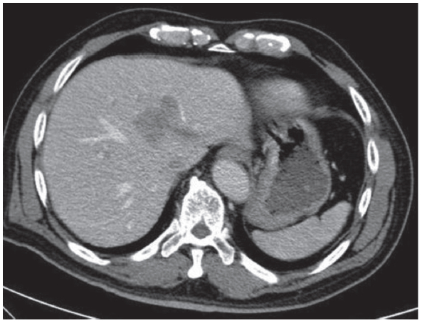
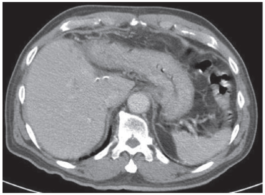
Figure 1:
The CT of abdomen showed an ill-defined hypodense mass with neovasculature stain at liver segment 3, measuring 4.5 x 3.5 x 2.9 cm.
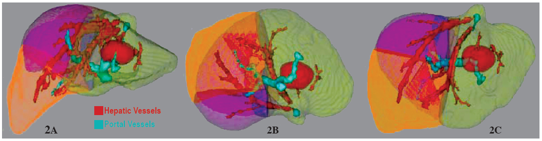
Figure 2A-C:
The 3D reconstruction of liver with demonstrated the size of liver mass at left lobe in correlation to hepatic vein (red) and portal vein (green)
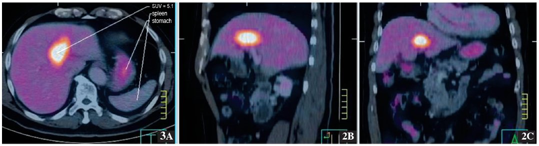
Figure 3A-C:
The PET/CT scan showed liver mass at segment 3 of liver with increasing metabolic activity SUV of 5.1.
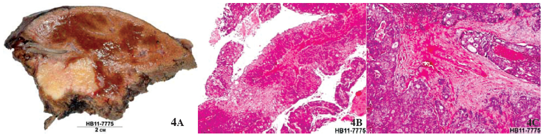
Figure 4A-C:
The specimen (4A) showed a liver mass. The microscopic examination (4D-C) revealed adenocarcinoma.
Figure 5:
The CT of abdomen with contrast enhancement
revealed status post left hepatectomy, No evidence of tumor recurrence at surgical margin is observed. IVC is intact.
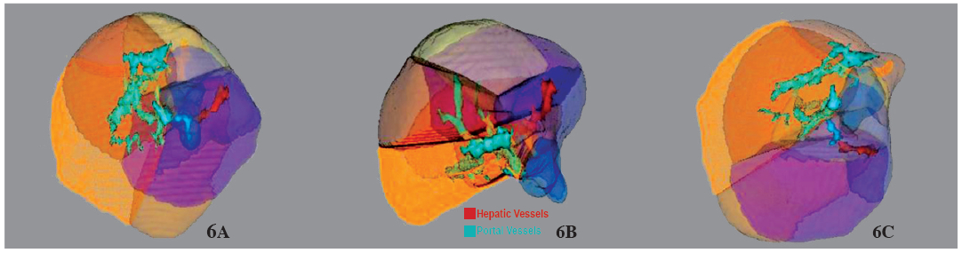
Figure 6A-C:
The 3D reconstruction of liver post left hepatectomy, patency of hepatic vein (red) and portal vein (green) are seen.