Electronic ISSN 2287-0237
Chemotherapy and radiotherapy,separately or sequentially, are established protocols in the management of lung malignancy. However, as early as 1999 a study in Osaka, Japan concluded that the concurrent approach yielded a significantly increased response rate and enhanced median survival duration when compared with the sequential approach [as applied to selected patients with unresectable stage III non-small cell lung cancer (NSCLC)].1
Several other studies and clinical trials that were subsequently published validated the efficacy of concomitant chemotherapy and radiotherapy in the management of NSCLC.2
While previous studies did indeed advance novel treatment methods for NSCLC, the team in Bangkok Hospital found it curious that even in a recent study published in April 2012,3 the most advanced staging evaluation methods used were mediastinoscopy or mediastinotomy (following bronchoscopy, bone scan, computed tomography (CT) scan of the chest) to acquire biopsy samples from lymph nodes. A randomized trial in 2010 of two-hundred and forty one patients revealed that a staging strategy combining endosono- graphy and surgical staging showed a higher sensitivity rate for mediastinal nodal metastases when compared to surgical staging alone among patients with (suspected) NSCLC.4 A minimally- invasive procedure, endobronchial ultrasonography is currently considered the gold standard in the evaluation of mediastinal lymph nodes and lung lesions along with its other usage in the clinical set-up.
We designed a study that will explore the efficacy of concomitant use of chemotherapy and radiotherapy by using endobronchial ultrasound (a low-risk method), as an evaluation tool for downstaging of lung cancer patients not limited to NSCLC.
Eligibility Criteria
The patients who were selected for this study were histologically or cytologically confirmed as newly diagnosed and untreated for lung cancer. They were aged 18 and above, without intake of aspirin or any blood thinners 7 days prior to procedure and signed a written consent for treatment in the hospital and for receiving the EBUS procedure in the operating room.
Exclusion Criteria
Patients who were excluded were pregnant or nursing, or fertile patients who aren’t using effective contraception; had previous thoracic radiotherapy, chemotherapy, immunotherapy or biologic therapy for lung cancer; or blood thinner intake 7 days prior to procedure.
Endosonography
A combination of endobronchial ultrasound-guided transbronchial needle aspiration (EBUS-TBNA) and endobronchial ultrasound with guide sheath (EBUS-GS) was used to obtain sample specimens for biopsy. During endobronchial ultrasound (EBUS) procedures, the lungs and mediastinum were visualized for possible pathology or lesions through a flexible scope. Ultrasonic waves enabled the operator to see structures through the wall of the airway. Samples of the lymph nodes and any masses were taken as necessary. Biopsy specimens underwent On-site pathology/Rapid on-site evaluation (ROSE). The remaining tissue samples were sent to the laboratory for further cytological and immunohistochemistry testing. Tumor gene mutation deficiencies were also investigated via Anaplastic lymphoma kinase (ALK) and the epidermal growth factor receptor (EGFR) tests.
Chemotherapy and Radiotherapy
The patients with definite diagnosis of lung cancer were referred to oncologists who prescribed patient-specific chemotherapy and radiotherapy drug combination.
A 72-year-old male patient was previously being treated for chronic obstructive pulmonary disease (COPD) when chest x-ray revealed left lower lung mass. Carcinoembryonic antigen (CEA) was 108.90ng/ml and positron emission tomography and computed tomography (PET/CT) Scan showed there was a 3.5x4.0cm speculated mass in the superior segment of the left lower lobe with radiating strands to the hilum and the fissure. Another tiny subpleural nodule was present at the posterior gutter of the left lower lobe. There are a few 0.6-0.9cm nodes at the interlobar station of the left hilum. There was another 1.0cm pleural based nodule at the posterior segment of the right upper lobe with retraction of the fissure. No pleural effusion was seen. There are a few 0.5-0.7cm nodes in the lower paratracheal and AP window regions. The finding of positron emission tomography (PET) scan in the thorax was a left lower lobe lung mass showing markedly increased the foundations of digital Games (FDG) uptake with maximum standardized uptake value (SUV) of 6.1. Hypermetabolic hilar node was also noted with SUV of Faint metabolic activity of nodule in right upper lobe and right hilar region was noted with SUV of 0.8 and 1.1 respectively. Bronchoscopy and biopsy were done which revealed small sheets of tumor cells found with highly pleomorphic enlarged hyperchromatic nuclei, occasional small nucleoli and abundant vacuolated cytoplasm. He was then diagnosed of non-small cell carcinoma.
The patient underwent sessions of radiation and chemotherapy treatment. He was able to tolerate the medications and therapy until 6 months later, when he developed left lung atelectasis. His chest x-ray showed retrocardiac left lower lobe atelectasis appearing slightly increased. Small amounts of bilateral pleural effusion, more on the left side were noted. The CT scan of chest revealed right pneumothorax, measuring about 0.7 cm in thickness. Pulmonary infiltration, more on the superior segment of left lower lobe, where atelectasis was increased. Fluid in left bronchi was observed.
Fiberoptic bronchoscopy was done and revealed endo- bronchial obstruction of the medial and lateral segment of left lower lobe. A tumor at the medial segment was completely removed but the lateral segment tumor was hard as stone. Biopsy was done instead and bleeding was managed by electrocautery. Cytopathology report was acute and chronic bronchitis with mild squamous metaplasia. Necrotic tissue with fibrinopurulent exudate was also found. Microscopic investigation showed that two pieces of necrotic tissue, with fibrinous material and areas of acute inflammation and one cluster of a few tiny fragments of bronchial mucosa showing mild acute and chronic inflammation with areas of mild squamous metaplasia. No epithelioid granuloma or malignancies were observed.
He was then treated for atelectasis and bronchitis. The cytopathology results from bronchoscopy confirmed no recurrence of lung cancer.
A 60-year-old male, was diagnosed with limited small cell lung cancer post chemo, thoracic radiation therapy and prophylactic whole brain radiation therapy (WBRT). His last dose was given 3 months prior to consultation.
Three months prior to consult, the patient complained of dysphagia, body weight loss of 8 kilograms (kg) for the past 2 months, chest discomfort that radiates to the abdomen and voice hoarseness.
Physical examination revealed that he appeared thin, had diminished breath sounds upon auscultation, no coughing, no sputum, and had no fever episodes. PET/ CT scan revealed there is a 3.0x3.5cm speculated mass with scattered punctuate calcifications at the posterior segment of the right upper lobe with extension to the hilum, broad attachment to the fissure and volume loss of the right hemithorax (Figure 1). There are scattered foci of reticulonodular opacities in the upper lobe, superior segment right lower lobe and the right middle lobe, possibly representing underlying or superimposed infection. A few nonspecific tiny subpleural nodules were also found in both lungs. There was no pleural effusion. A 0.7cm right hilar node and 0.6cm subcarinal and right lower paratracheal nodes were observed. PET scan revealed speculated mass in the posterior segment of right upper lung and showed faint uptake of the FDG with SUV of 1.3. Opacities in right upper and right lower lung also showed minimally increased FDG uptake with SUV of 1.6. The metabolic activity of each hilum or mediastinum was not increased. EBUS-GS and EBUS-TBNA were done to investigate if there was evidence of recurrence of lung cancer.
Brushing slide specimen revealed therapy-induced atypia and mediastinal lymph node was negative for granuloma and malignancy. Fluid and brush showed benign reactive bronchial cells along with some foamy macrophages, lymphocytes and neutrophils. Some atypical cells were seen with enlarged cells, vacuolated cytoplasm, multi- nucleated nuclei and prominent nucleoli. There was no evidence of granuloma or malignancy. Smears contain mature lymphocytes which have small dark nuclei and scanty cytoplasm. The polymorphous population of lymphocytes was also seen. The background shows numerous red cells and benign bronchial epithelial cells. There was no evidence of tumor or granuloma. Bronchial biopsy revealed hyalinized fibrosis, no evidence of granu- loma or malignancy.
With the provided results, the tumor panel concluded at that moment, there was no evidence of recurrence of small cell lung cancer.With the provided results, the tumor panel concluded at that moment, there was no evidence of recurrence of small cell lung cancer.With the provided results, the tumor panel concluded at that moment, there was no evidence of recurrence of small cell lung cancer.
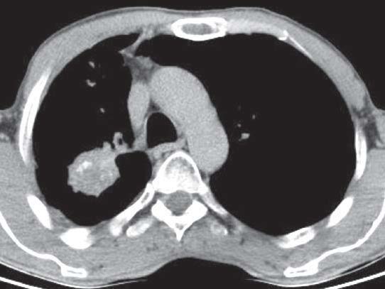
Figure 1: A picture of the CT scan result as described in case study # 2
A 50-year-old male caucasian patient was diagnosed with adenocarcinoma stage II. He was a former smoker for 6 years and has family history of lung cancer.
Three months prior to consultation, he developed fever, cough, anorexia, body weight loss of 13kg and pain in with his bones. He was then diagnosed with giardiasis and gastritis but was subsequently treated. He regained his appetite but his cough still persisted. One month prior to consultation, he had had several episodes of febrile (temperature 39.8oC), sweating, and cough. He went for a medical consultation in Chiang Mai, Thailand where he was tested for tuberculosis which came back negative. CT scan of chest revealed mass at the right upper lobe with mediastinal node enlargement.
Initial assessment revealed diminished lung breath sounds at right upper lobe lung. PET/CT scan was done and revealed an irregularly outlined soft tissue mass at the posterior segment of the right upper lobe with a broad attachment to the fissure. The mass measures about 8.2x4.0 cm. There was central necrosis at the main component of the mass and cavitation at the peripheral aspect, and at the medial extension behind the right main bronchus. There was no abnormality in the rest of the lungs or pleural effusion. A 0.9cm node was seen at the precarinal space and a few 0.5-0.7cm nodes in the right lower paratracheal space. PET scan findings of density in the right upper lung showed increased FDG uptake with SUV max of 5.3. The cavitating postbronchial component showed a hypermeta- bolic activity with SUV of 1.8. CEA was 1.70ng/mL and Quantiferon TB result was negative.
EBUS-GS and EBUS-TBNA were advised for further assessment and evaluation. The onsite pathologist revealed that the specimen obtained from the upper lobe mass revealed adenocarcinoma. However, the two slides from right paratracheal nodes were negative for malignancy. Post operatively, the patient did not manifest pneumothorax, bleeding, and infection.
The official cytopathology report revealed that the first smear contained mature lymphocytes which have small dark nuclei and scanty cytoplasm. The polymorphous population of lymphocytes was also seen. The background showed numerous benign bronchial epithelial cells. The second, showed more bloody background. There is no evidence of tumor or granuloma. The second smear contains some clusters of atypical cells which have enlarged vesicular nuclei, prominent nucleoli and foamy cytoplasm. The background showed benign bronchial epithelium and numerous neutrophils. Bronchial biopsy revealed a section of a small piece infiltrated of malignant glandular epithelium with nuclear enlargement, hyperchromicity, small nucleoli and moderate amount of pink foamy cytoplasm. The larger piece showed only benign bronchial mucosa with numerous neutrophils and fibrin material. Tumor mutation from EGFR and ALK results were both negative.
In cooperation with Dr. Lodi, the patient was treated under two protocols. First, was the combination of Taxotere 10mg and Carboplatin 50mg alternating every other week with the second protocol was Cisplatin 10mg, Eteposide 10mg and Vinorelbine 10mg.
After the treatment, the patient decided to go back to his home country, underwent lobectomy which revealed no presence of malignancy in lung tissue.
Endoscopic ultrasound-guided fine needle aspiration (EUS-FNA) is a mediastinal staging modality that has been developed in the last decade. High sensitivities and speci- ficities have been reported in a large number of studies.4
The procedure is well tolerated, safe and has a high diagnostic accuracy (89-95%) for the analysis of mediastinal lymph node (LN). So far, no complications of EUS-FNA in the analysis of mediastinal LN have been reported. The advantages of this technique are multiple: tissue samples are obtained (in contrast to the imaging technique of CT) and the procedure itself is minimally invasive, is performed in an out-patient setting and can be repeated a number of times without technical difficulties (in contrast to mediastinoscopy).5
A similar study published in 2008 investigated the accuracy and sensitivity of EBUS-TBNA for restaging the mediastinum after induction chemotherapy in patients with NSCLC. The team from the University of Heidelberg concluded that despite EBUS-TBNA being a sensitive, specific, accurate, and minimally invasive test for mediastinal restaging of patients with NSCLC, tumor-negative findings should still be confirmed by surgical staging before thoracotomy.6
A study in Japan, on the other hand, stated that for mediastinal staging in lung cancer, the diagnostic yield of EBUS-TBNA is comparable to surgical staging in patients with enlarged lymph nodes.7
The above studies are important contributions to the development of EBUS. However, their research does not include analysis and evaluation of the downstaging of lung cancer after combined chemotherapy and radiation treatment. To our knowledge, this study in Bangkok Hospital is the pioneering report with which combined EBUS-TBNA and EBUS-GS has been used as an evalua- tion tool to measure downstaging of lung cancer after combined chemotherapy and radiation treatment.
Endobronchial Ultrasonography (EBUS) is an effective tool in evaluating downstaging in patients with lung cancer after combined chemotherapy and radiotherapy.
EBUS-GS provides a pathway to peripheral pulmonary lesions, enabling the operator to obtain short-axis bronchial views. It is a useful method for collecting samples from peripheral pulmonary lesions, including those which are too small to be visualized under fluoroscopy. The use of EBUS-TBNA is known for increasing diagnostic yield in obtaining a specimen for biopsy. It can be used for collecting both cellular and tissue specimen collection. With this capability, administering these procedures in tandem can effectively diagnose lung cancer as well as other diseases such as sarcoidosis and malignant lymphoma. The two procedures, complementing each other in many ways, are both safe for the patient: minimally invasive, relatively low risk and avoiding possible unnecessary thoracotomies.8


