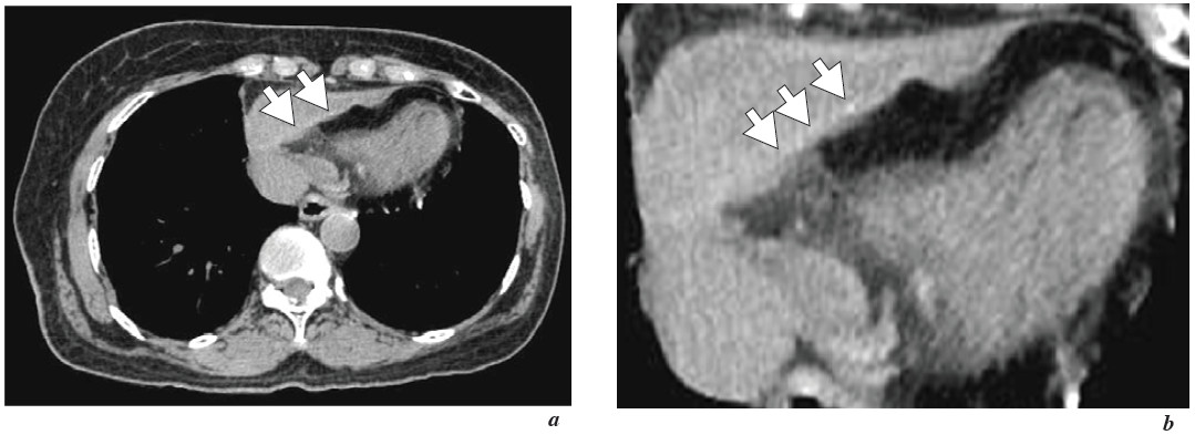
Figure 1 a-b: CT Chest shows thick pericardium at anteroinferior border with multiple foci microcalcification at wall of pericardium. This indicats granulomatous condition. Tuberculous pericarditis should be considered.
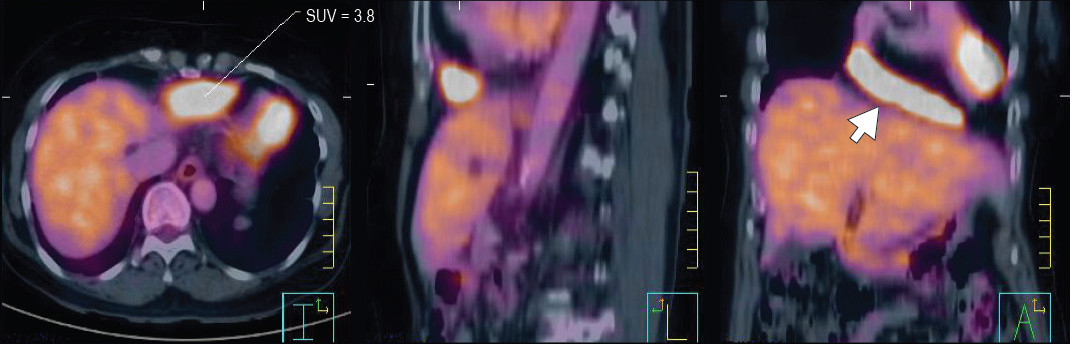
Figure 2: The 18FDG PET/CT scan shows increased metabolic activity activity at pericardium (SUV = 3.8).
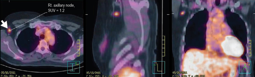
Figure 3: The 18FDG PET/CT scan shows increased 18FDG uptake at right axillary node (SUV = 1.2).
Figure 4: The tuberculin skin test shows positive result.
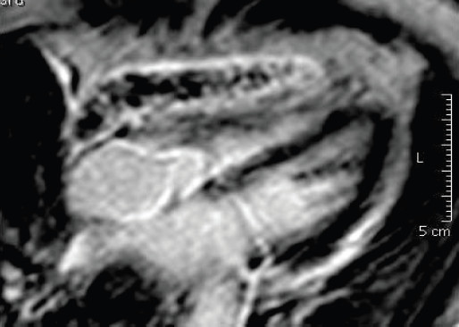
Figure 5: The MRI study of the heart with gadolinium shows localized thick wall of pericardium with rim contrast enhancement.
The inner wall of pericardium was thickening and irregular fine filling defects projected into the pericardial effusion which is compatible with chronic granulomatous pericarditis.
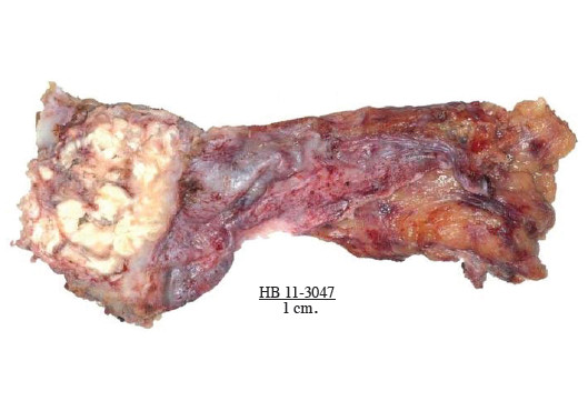
Figure 6: The gross specimen consisted of a fibrocalcific mass size 3.5x3.5x1.8 cm.
Cut section of the mass showed old and recent caseous material. The pericardium close to the mass was scarred and thick, up to 0.8 cm. The pericardium away from the mass was relatively normal.