Electronic ISSN 2287-0237
Digital tomosynthesis (DT) has been well described as a technique and the advantage of this procedure has been discussed previously.1 The most recognized technique is digital breast tomosynthesis (DBT).2 We focus here on different medical areas such as the chest, skeletal system, and urolithiasis among others. We will present a few cases using DT in assisting in the case of doubtful radiography and /or computed tomography (CT) image in other medical fields excluding DBT.
Digital Tomosynthesis in Chest Examination
A 61-year-old Thai female at a general checkup, had a chest radiograph that reveals a suspected faint density at the left lower lung field; its nature may be either a nipple or lung parenchymal lesion (Figure 1A). Chest tomosynthesis reveals a small pulmonary nodule 4mm in diameter at the peripheral region at the left lower lobe (Figure 1B), and the CT image confirmed this lesion (Figure 1C).
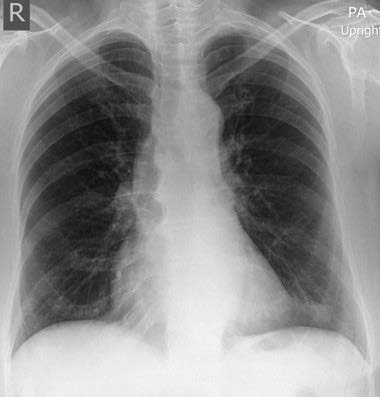
Figures 1A: Chest radiograph reveals localized haziness at the left lower lung field, nature is undetermined.
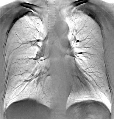
Figure 1B: DT reveals subtle pulmonary nodule 4 mm in diameter at left lower lobe.
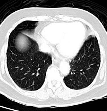
Figure 1C: CT image reveals small pulmonary nodule identical to DT finding.
A 54-year-old Thai male, undergoes a protocol chest CT for lung cancer detection. The high resolution CT image shows a subtle parenchymal pulmonary nodule with a spicular appearance at the right middle lung field. A DMT is requested to determine its nature (Figure 2A). DT reveals no evidence of any pulmonary nodule. The lesion at the arrow is at the junction of the pulmonary artery and the pulmonary vein crossing each other which simulates a pulmonary nodule (Figure 2B). DT is helpful in cases where a chest CT is suspicious.
James T. Dobbins III and colleagues’ studied3 156 nodules identified by CT; the detection sensitivity was percent for chest radiography and 13.4 percent for DT. The International Association for the study of Lung Cancer (IASLC)4 concluded that the use of DT in the early detection of lung cancer is comparable to the rates of low dosage CT and may offer an alternative to CT screening. It has promise to become a first-line lung cancer screening tool. Furthermore, DT has a lower cost and lower radiation dosage.
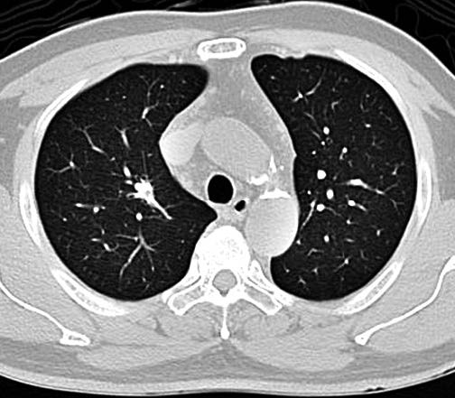
Figure 2A: CT image reveals subtle pulmonary nodule with spicular border see arrow.
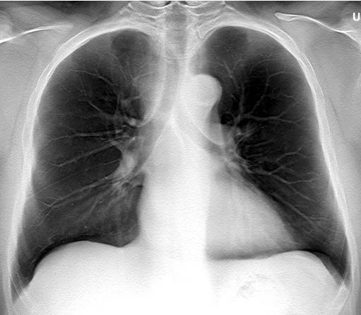
Figure 2B: DT reveals no pulmonary mass, the finding reveals the structures of pulmonary artery and vein across each other simulating nodule.
Digital Tomosynthesis in Skeletal System
A 65-year-old Thai male presents with swelling with redness and tenderness at the right sternoclavicular region for 3 weeks after clinical diagnosis of arthritis and local injection of steroid drug. The patient still complained of local tenderness. Conventional radiography at this region revealed no abnormality. DT at the right sternoclavicular region reveals a definite linear fracture at the right 1st cartilage (see arrows (Figure 3)). He recovered over time with conservative treatment.
Figure 3: DT at right sternoclavicular joint reveals definite evidence of linear fracture at the cartilage of the right 1st rib.
A 34-year-old Thai male presented with pelvic pain after a car accident. CT image reveals a small piece fracture at the right pubic ramus (Figure 4B-C). DT pelvis reveals a comminuted fracture at the right pubic ramus (Figure 4D). The image shows a more delicate and precise rendition than a CT image. Gothlin JH and Geijer M studied5 DTs of total hip joint arthroplasty with a suspicion of loosening in 40 patients. DT demonstrated improved vsualization of demineralization and the extent of osteolysis and demar- cation when compared to radiography. The skeletal digital tomosynthesis may detect conditions more precisely than radiography. Takatoshi Aoki, et al6 studied 20 cases of patients with an established diagnosis of rheumatoid arthritis. They compared the results from radiography, tomosynthesis and MRI of the hand and wrist joint base for bone erosion. The results of the detection rates were 26.5%, 36.1% and 36.7% respectively. The rate of detection when using tomosynthesis is superior to radiography and almost comparable to MRI, furthermore, the radiation level for radiography is 0.13 mGy and 0.25 mGy for tomosynthesis.
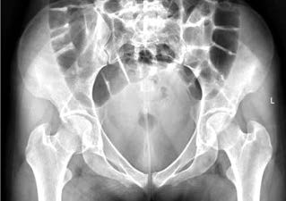
Figure 4A: Pelvic radiograph reveals suspicious fracture at right superior pubic ramus.
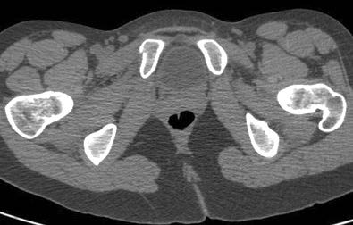
Figure 4B: CT image on axial section reveals small area of fracture at right superior pubic ramus medially.
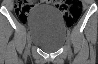
Figure 4C: CT image on coronal section reveals minor linear fracture proximal to the right superior pubic ramus.
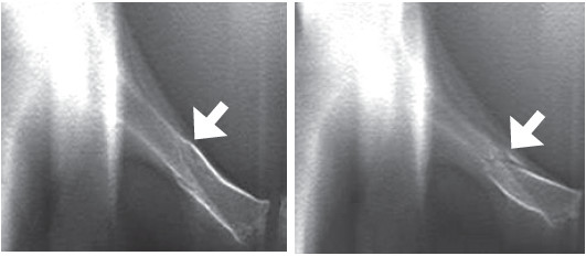
Figure 4D: DT reveals definite evidence of comminuted fracture at mid portion of the right superior pubic ramus.
Digital Tomosynthesis in the Urologic System
A 60-year-old Thai male presented with sudden abdominal pain, the KUB radiograph reveal a suspected small opaque left renal stone vs. feces see arrow (Figure 5A). DT reveals definite evidence of an opaque stone at the left kidney (Figure 5B).
The diagnosis of urolithiasis is normally confirmed by conventional radiography but recently unenhanced CT images have been found to be superior in the precise identification of the stone as well as its location.7 This modality reflects that DT should be the same quality as an unenhanced CT image.
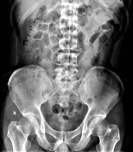
Figure 5A: The KUB radiograph reveals a small density at left lumbar region, which may due to a residual renal stone or fecal material.
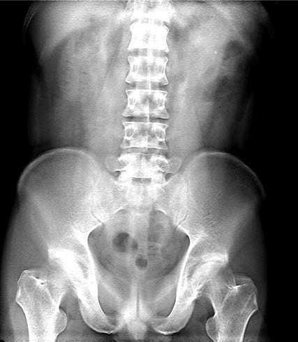
Figure 5B: The DT reveals definite evidence of a small opaque stone at the left kidney.
Digital tomosynthesis applies to almost every system in the body including breast, chest, musculoskeletal, skeletal metabolic disorders such as rheumatoid arthritis and disorders in the urologic system. This modality is very helpful in assisting the detection of pathology with low cost, low radiation dosages and a fast examination time. We introduced a few samples of cases presenting with various problems in multiple medical fields for its advantages to the radiologist, general physician, thoracic physician, urologist, rheumatologist, radiographic techni- cian and patients who are interested in new technology and continuing education. It is expected that this technique for routine examination will be updated when this modality is suitable for the patient’s requirements.
Digital tomosynthesis (DT) demonstrates that it is another imaging modality (excluding digital breast tomography (DBT)) which is useful in detecting lung cancer or other lesions in the chest, for examining the skeletal system in cases of fracture or articular surface of joints (such as hip joint or soft tissue mass) or in the detection of urolithiasis. DT is less costly, has lower radiation rates and is a fast examination. We recommend DT to assist in the detection of these entities as described.


