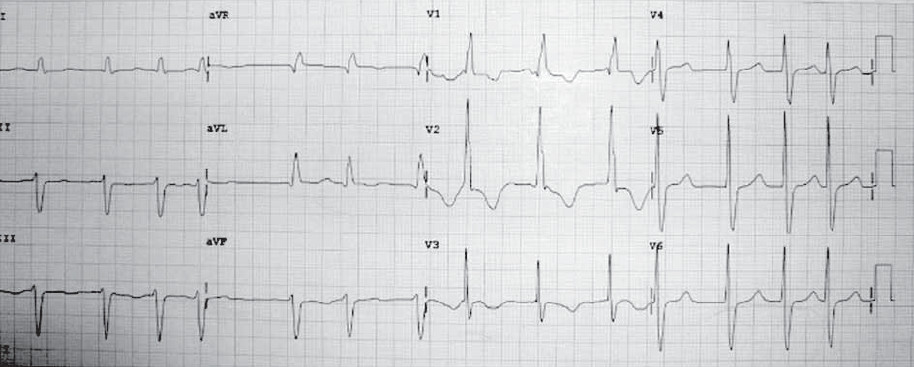
Figure 1: ECG on admission showed atrial fibrillation, RBBB with secondary ST-T changes.
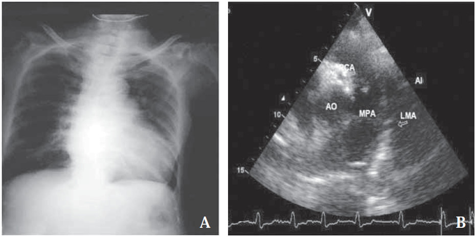
Figure 2: Left: Chest film five months before this event showed cardiomegaly and prominent pulmonary trunk (A). Right: short axis view of the aortic root and pulmonary trunk that showed origin of RCA and LMA.
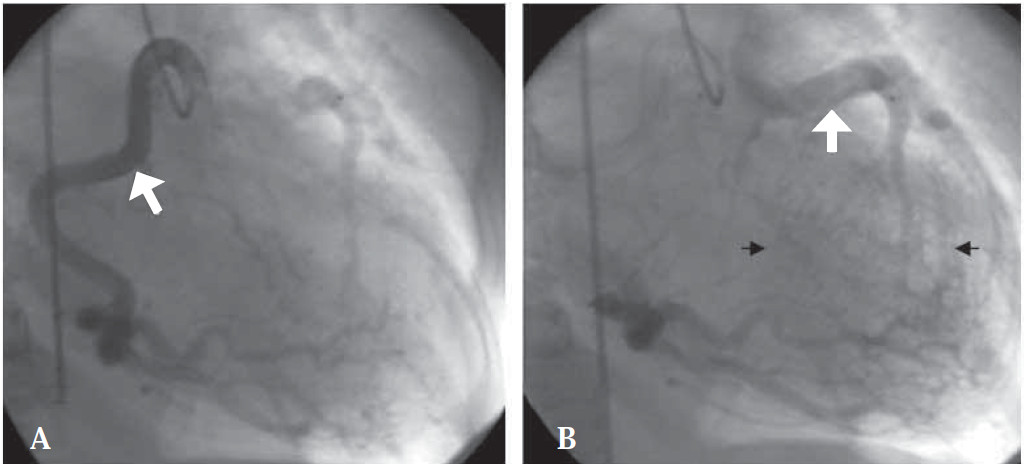
Figure 3: Left heart CAG images revealed (A) the RCA which was in the right position (A) with no show of the left coronary artery origin at the left cusp sinus ,
(B) the left coronary arteries (white arrow) were supplied by the right to left collaterals (black arrow).
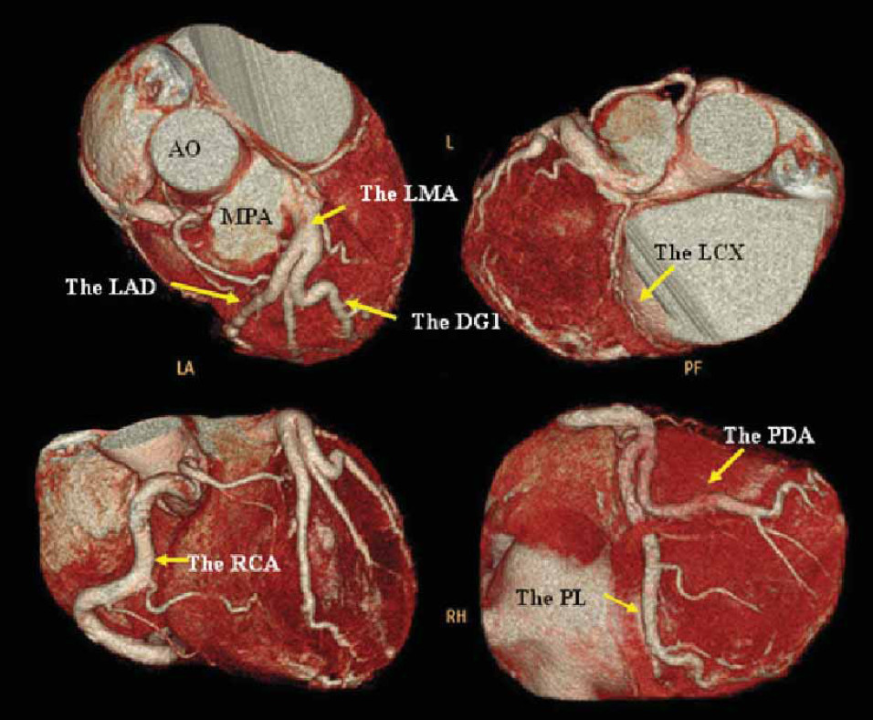
Figure 4: Volume rendering images of the 256- slice MDCT revealed the large RCA which originated from the right cusp and the LMA originated from the MPA (ALCAPA).
AO= Aorta,
MPA= Main pulmonary artery,
LMA = Left main artery,
LAD = Left descending coronary artery,
LCX= Left circumflex coronary artery,
RCA = Right coronary artery,
PL= Posterior lateral artery,
PDA = Posterior descending artery