Electronic ISSN 2287-0237
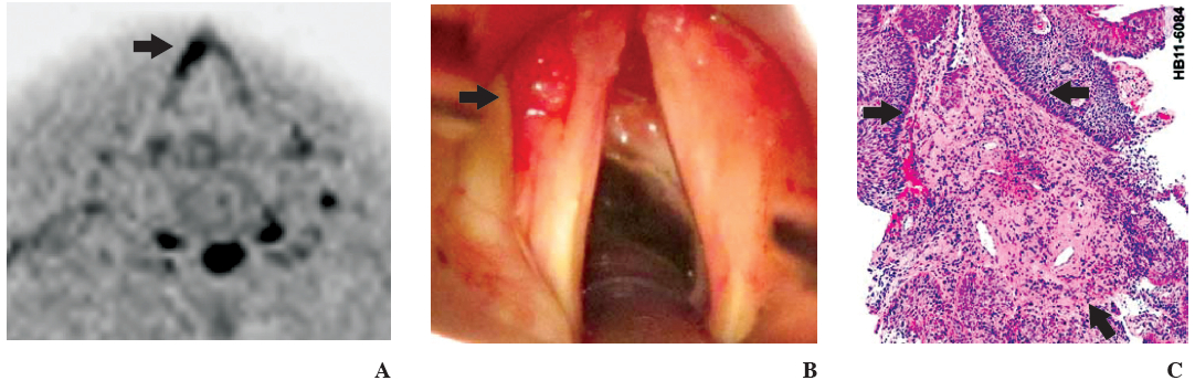
A79-year-old man man presented with hoarseness since 3 months, he was a heavy smoker for many years. Indirect laryngoscopy was unable to demonstrate the lesion. MRI-Diffusion Weighted pulse sequence at vocal cord (Figure A) revealed localized hyper intensity (reversed image) at anterior one third of left vocal cord.1 The patient underwent general anesthesia. Then a direct laryngoscopy revealed the tumor. Exoplytic mass at anterior haft of left vocal cord is shown in Figure B. Anterior commissure appears intact. The microscopic examination (Figure C) revealed neoplastic proliferation of squamous epithelium of larynx with atypia. There was evidence of early invasion into subepithelial tissue.
This example illustrates the usefulness of DW pulse sequences, which can make abnormalities in certain pathologies more obvious,especially for identifying small tumors at the vocal cord, as in our case.


