Electronic ISSN 2287-0237
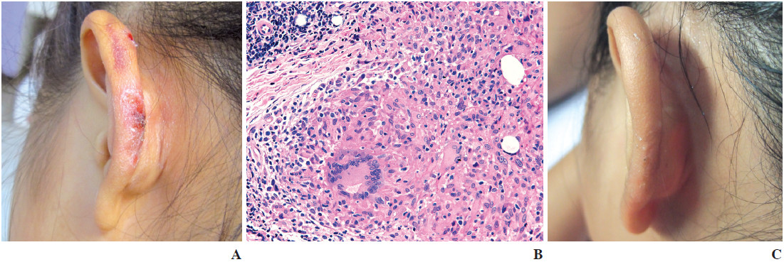
A 9-year-old Thai girl presented with pink plaques on the left ear for 6 months without any symptoms. She received topical steroids and antibiotic without clinical improvement. Physical examination revealed well demarcated, irregularly bordered, pink infi ltrated plaques on the left ear (Figure A). Apple-jelly color was seen when examined by diascopy. No regional lymphadenopathy was detected. Other systemic examinations were unremarkable. The histopathological examination revealed tuberculoid granuloma with Langerhans giant cells in the papillary dermis (Figure B) which was a hallmark of cutaneous tuberculosis. The tissue was negative for acid-fast bacilli, polymerase chain reaction and mycobacterium culture. The purifi ed protein derivative (PPD) Mantoux test was positive with 20mm induration. Chest x-ray was unremarkable. She received anti-tuberculous treatment as standard regimen (2 HRZE/4HR). She was able to tolerate the medications well and showed marked clinical improvement after treatment (Figure C).
Lupus vulgaris is the most common form of cutaneous tuberculosis. Chronic progressive plaque is a typical characteristic. Lupus vulgaris can be acquired as result of direct extension through lymphatic or hematogenous spread or rarely by cutaneous inoculation of M. tuberculosis. It most often develops, following cervical lymphadenitis or pulmonary tuberculosis and the most common sites are nose and cheek.1
Lupus vulgaris can be diagnosed mainly by clinical and histopathological features. Standard anti-tuberculous medications are very effective and well tolerated.1-2


