Electronic ISSN 2287-0237

VOLUME

A CASE OF LUNG CARCINOID TUMOR, SUCCESSFULLY TREATED WITH BRONCHOSCOPIC CRYOSURGERY IN THAILAND

SEPTEMBER 2015 - VOL.10 | CASE REPORT

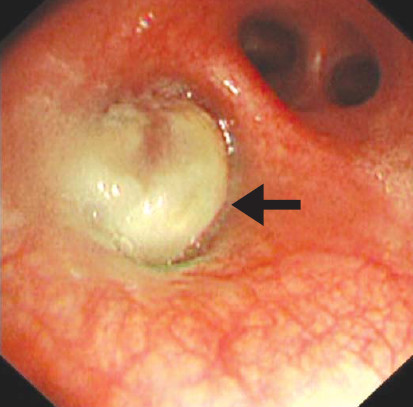
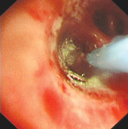
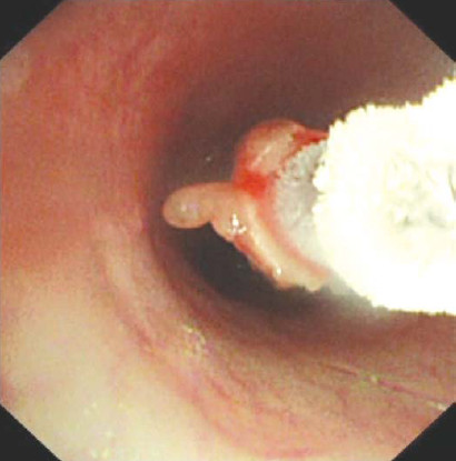
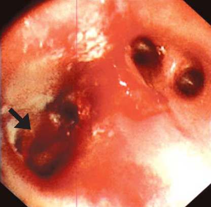
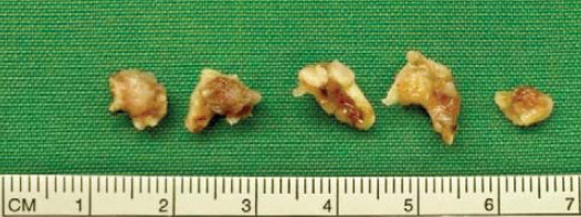
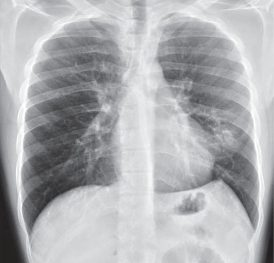
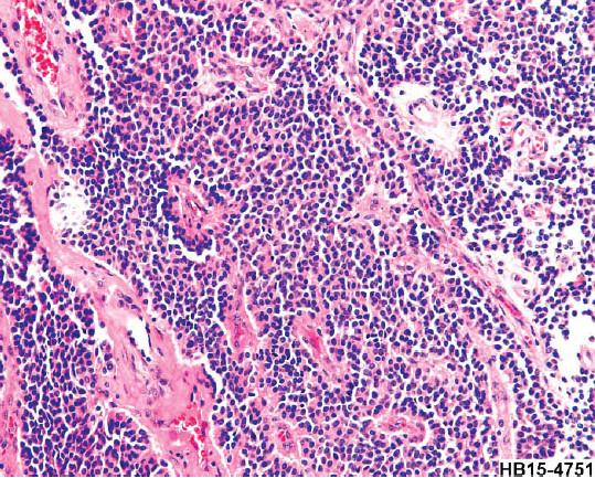
A lung carcinoid tumor is a rare malignant neoplasm. The clinical presentation depends on the characteristics of the tumor, its location and hormones secreted. The most common symptoms are cough, wheezing, chest pain, hemoptysis and dyspnea. Diagnosis can be established through radiographic images, biopsy of the tumor with tissue analysis supplemented by blood tests. In previous years, surgery has been the gold standard in treating carcinoid tumors. However, recently, there have been several studies conducted in the efficacy of the use of bronchoscopic methods such as cryosurgery in completely extracting and treating lung carcinoid tumors without metastases. This case report’s objective is to illustrate that cryosurgery should be considered as the new gold standard in treating lung carcinoid tumors.

Keywords

cryosurgery, cryotherapy, lung carcinoid tumor, endobronchial tumor, argon plasma coagulation

DOI:

10.31524/bkkmedj.2015.09.006

MEDIA
Figure 1: The CT scan image illustrates the carcinoid tumor at the left upper lobe lung (arrow with obstructive atelectasis).
Figure 2: 18F-FDG PET/CT reveals hypermabolic focus at lingular segment of left upper lobe, the 18F-FDG uptake SUV of 1.9.
Table 1. This shows the patient’s lung function test prior and post cryosurgery. The FEF25-75% has significantly increased from 75% to 89%.
Figure 2: The bronchial tumor as viewed through the virtual bronchoscopy.
Figure 3: The carcinoid tumor as directly viewed from the bronchoscope.
Figure 4: The carcinoid tumor when carbon dioxide is applied.
Figure 5: The frozen carcinoid tumor being pulled out from the bronchus.
Figure 6: The lung parenchyma post cryosurgery with minimal bleeding.
Figure 7: The carcinoid tumor extracted by pieces through cryosurgery.
Figure 8: The chest x-ray result shows localized patchy infiltration at the lingular segment of the left upper lobe.
Figure 9: Tumor cells in organoid and trabecular pattern in hyalinizing stroma with some palisading at periphery. The tumor cells have fi ne granular tissue chromatin, indistinct nucleoli. The findings are compatible with atypical carcinoid.
SEARCH VOLUME & NUMBER
GO
RECENT VOLUME & NUMBER